How to Videos
Biopsy Collections
Skin biopsies are essential procedures for health care providers who manage skin conditions. These office-based procedures can be used for the diagnosis of suspicious dermatologic lesions or definitive treatment for some malignant, irritated, or precancerous lesions.
This video will walk you through best practices for performing cutaneous biopsies.
Necesssary Biopsy Supplies
Proper exam room setup is essential for reducing prep time and improving efficiency when performing biopsies.
This educational video teaches the recommended tools needed for each type of biopsy.
Supplies covered in this video:
- Alcohol Prep Pads
- Syringes & 21 Gauge Needles
- Cap & Rubber Guard
- 3 & 4mm Biopsy Punch
- Forceps
- Formalin Fixatures
- Scalpel with Attached 15 Blade
- Split Double-edged Razor Blades
- Iris Scissors
- Michels Fixatives
- Zamboni Fixatives
- Derma Blades with Plastic Guards
Browse Types of Biopsy Videos
Punch Biopsy
Punch biopsies yield full-thickness samples and can be used for diagnosing lesions that require dermal and subcutaneous tissue. They are ideal for tumors that are too deep to be sampled by shave technique and for inflammatory conditions that have a deep component. Punch biopsies smaller than 3mm should be avoided.
Collection Guides
Superficial Shave Biopsy
The shave biopsy technique is commonly used because of how quickly it can be performed, the simplicity of wound care, cosmesis, and cost-effectiveness.
A superficial shave biopsy removes a thin disk of tissue typically by scalpel (generally a #15 blade), although most physicians prefer a double-edged razor blade. The procedure yields a flat, thin specimen of combination epidermis and upper dermis.
As such, it is the most effective method of sampling raised lesions such as warts, papillomas, skin tags, pyogenic granulomas, non-melanoma skin cancers, and a variety of keratoses.
Saucerization Biopsy
In a saucerization biopsy, a thick disk of tissue is removed with a curved blade yielding a specimen that extends to at least the mid-dermis.
It is useful for sampling or removing larger lesions.
Wound/Lesion Swab
Although most lower extremity foot and leg ulcers may be venous, arterial, or neuropathic, patients presenting with a lower extremity nonhealing wound of long duration, which may not be responding to treatment (2 months or longer), should be considered for a wound biopsy (punch or curettage) procedure to determine the presence or absence of malignancy or inflammatory condition.
Ideally, two biopsies should be performed to include both the base of the ulcer (for malignancy) and the wound margin (for inflammatory causes).
Collection Guide
Related Video
Fine Needle Aspiration Biopsy
A fine needle aspiration biopsy provides a sample of fluid and/or cells from a deep-seated subcutaneous mass for diagnosis.
Avoid removing a soft tissue mass before determining a definitive diagnosis based on the fine needle aspiration biopsy, such as lipoma, ganglion cyst, sarcoma, soft tissue tumors, or lesions that may resemble epidermal inclusion cysts.
Uncapped Needle? Watch this syringe re-capping video to see how safely re-cap a needle using the one-handed scoop technique.
Collection Guide
Related Video
Epidermal Nerve Fiber Density Biopsy Procedure
In this instructional video, Lilly Khavari, DPM, a board-certified foot and ankle specialist, demonstrates the precise technique for collecting an Epidermal Nerve Fiber Density (ENFD) skin punch biopsy.
The ENFD biopsy is the clinical gold standard for diagnosing Small Fiber Neuropathy (SFN), a condition commonly presenting with symptoms such as burning, prickling, numbness, or shooting pain. Accurate specimen collection is critical to obtaining reliable diagnostic results.
Collection Guide
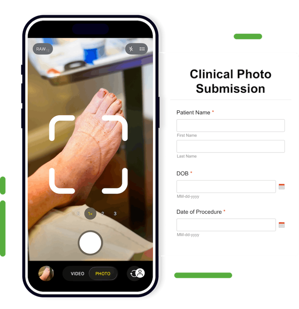
Securely Submit Clinical Photos
Our Clinical Photo Submission form makes it easy to securely and efficiently send clinical photos directly to Sagis from any device.
The form is HIPAA-compliant, encrypted, and designed for fast submission. You can capture a photo in the moment or upload an existing image from your library. Include measurements, a gross visual description of the lesion, and the patient’s clinical history in the visual description field.
Once submitted, your photo accompanies the case and is referenced in the resulting pathology report.
Bookmark the link to access the form from any cell phone, tablet, or desktop. QR code stickers for individual patient rooms are also available through your local Sagis representative or directly on our Clinical Supplies Order Form.
Biopsy Collection Videos
The 10 Commandments of Biopsy Collection
Skin biopsies are essential procedures for health care providers who manage skin conditions. These office-based procedures can be used for the diagnosis of suspicious dermatologic lesions or definitive treatment for some malignant, irritated, or precancerous lesions.
This video will walk you through best practices for performing cutaneous biopsies.
Biopsy Supplies and Set-up
Proper exam room setup is essential for reducing prep time and improving efficiency when performing biopsies.
This educational video teaches the recommended tools needed for each type of biopsy.
Punch Biopsy
Punch biopsies yield full-thickness samples and can be used for diagnosing lesions that require dermal and subcutaneous tissue. They are ideal for tumors that are too deep to be sampled by shave technique and for inflammatory conditions that have a deep component. Punch biopsies smaller than 3mm should be avoided.
Related Video

Superficial Shave Biopsy
The shave biopsy technique is commonly used because of how quickly it can be performed, the simplicity of wound care, cosmesis, and cost-effectiveness.
A superficial shave biopsy removes a thin disk of tissue typically by scalpel (generally a #15 blade), although most physicians prefer a double-edged razor blade. The procedure yields a flat, thin specimen of combination epidermis and upper dermis.
As such, it is the most effective method of sampling raised lesions such as warts, papillomas, skin tags, pyogenic granulomas, non-melanoma skin cancers, and a variety of keratoses.
Related Video
Saucerization Biopsy
In a saucerization biopsy, a thick disk of tissue is removed with a curved blade yielding a specimen that extends to at least the mid-dermis.
It is useful for sampling or removing larger lesions.
Related Video
Wound/Lesion Swab
Although most lower extremity foot and leg ulcers may be venous, arterial, or neuropathic, patients presenting with a lower extremity nonhealing wound of long duration, which may not be responding to treatment (2 months or longer), should be considered for a wound biopsy (punch or curettage) procedure to determine the presence or absence of malignancy or inflammatory condition.
Ideally, two biopsies should be performed to include both the base of the ulcer (for malignancy) and the wound margin (for inflammatory causes).
Related Video
Fine Needle Aspiration Biopsy
A fine needle aspiration biopsy provides a sample of fluid and/or cells from a deep-seated subcutaneous mass for diagnosis.
Avoid removing a soft tissue mass before determining a definitive diagnosis based on the fine needle aspiration biopsy, such as lipoma, ganglion cyst, sarcoma, soft tissue tumors, or lesions that may resemble epidermal inclusion cysts.
Uncapped Needle? Watch this syringe re-capping video to see how safely re-cap a needle using the one-handed scoop technique.
Related Video
Epidermal Nerve Fiber Density Biopsy Procedure
In this instructional video, Lilly Khavari, DPM, a board-certified foot and ankle specialist, demonstrates the precise technique for collecting an Epidermal Nerve Fiber Density (ENFD) skin punch biopsy.
The ENFD biopsy is the clinical gold standard for diagnosing Small Fiber Neuropathy (SFN), a condition commonly presenting with symptoms such as burning, prickling, numbness, or shooting pain. Accurate specimen collection is critical to obtaining reliable diagnostic results.