Presented by Jennifer Vickers, MD
Educational dermpath case series for dermatology residents
Patient: 27-year-old male
Lesion Location: Scrotum
A biopsy of a verrucous lesion on the scrotum was performed.
Take a moment to review the histologic images below, do you know how Dr. Vickers came to this diagnosis?
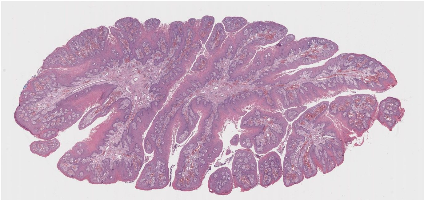
(Low-power histologic view of verrucous lesion)
On examination, several defining features stand out:
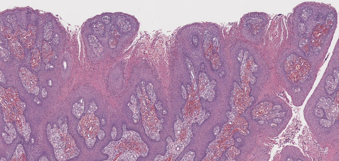
(Medium power showing papillomatosis)
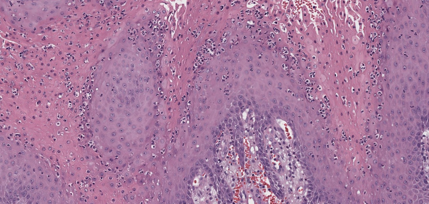
(High power showing parakeratosis and neutrophils)

(High power showing foamy macrophages in dermal papillae)
When evaluating a verrucous lesion like this, several differentials come to mind. Let’s walk through the three most likely contenders and highlight what sets them apart.
Etiology: Human papillomavirus (HPV), most commonly types 6 and 11
Histology:
Key Distinction:
Although condyloma and verruciform xanthoma both exhibit papillomatosis, koilocytes are the hallmark of condyloma. Verruciform xanthoma, in contrast, lacks HPV-associated changes.
Etiology: HPV (types 2 and 4 most common)
Histology:
Key Distinction:
While verruca vulgaris and verruciform xanthoma share a verrucous architecture, verruca vulgaris displays viral cytopathic effect and lacks the xanthomatous infiltrate that defines verruciform xanthoma.
Etiology: Low-grade variant of squamous cell carcinoma
Histology:
Key Distinction:
Unlike verruciform xanthoma, verrucous carcinoma shows invasive downward growth and a more aggressive epithelial proliferation. No foamy macrophages are seen.
The presence of foamy macrophages (lipid-laden histiocytes) within dermal papillae is diagnostic. This lesion is not associated with HPV, setting it apart from other verrucous lesions in the anogenital region.
📚 Quick Summary
| Feature | Verruciform Xanthoma | Condyloma Acuminatum | Verruca Vulgaris | Verrucous Carcinoma |
| HPV-related | ❌ No | ✅ Yes | ✅ Yes | ❌ No |
| Foam cells | ✅ Present | ❌ Absent | ❌ Absent | ❌ Absent |
| Koilocytosis | ❌ Absent | ✅ Present | ✅ Possible | ❌ Absent |
| Invasive growth | ❌ No | ❌ No | ❌ No | ✅ Yes |
| Behavior | Benign | Benign | Benign | Locally aggressive |
💬 Final Thought
Verruciform xanthoma is a classic dermatopathology “look-alike” that rewards careful microscopic examination. Remember: the foam cells tell the story.
Sagis Diagnostics is proud to support dermatology residents and dermatology residency programs through high-quality educational content and histopathologic learning resources.
Follow us on Instagram for more micro-learning opportunities with interactive cases and pathology insights.